

 InfoSearched
A cadaver lab reveals why a kidney stone can feel worse than childbirth or a gunshot wound.
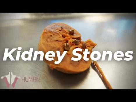
In “The Most PAINFUL Thing a Human Can Experience?? | Kidney Stones,” the Institute of Human Anatomy’s Justin Cottle pulls out real human specimens to answer a question most people only ask once they’re doubled over in an ER waiting room: why does a stone smaller than a pea produce pain severe enough to make grown adults vomit and beg for morphine. The answer, he explains, has almost nothing to do with the stone scraping tissue on its way out.
- Cottle demonstrates on cadaver specimens that the agony of renal colic comes primarily from a stone lodging in the ureter and backing up urine, not from the stone’s rough surface scratching tissue.
- The blockage triggers hydronephrosis — visible swelling of the kidney — along with violent muscular spasms of the ureter as it tries to force the obstruction through.
- Calcium oxalate stones are the most common type, followed by uric acid, struvite, and the rarer cystine stones, with dehydration, high sodium intake, and heavy animal protein consumption named as key risk factors.
The Anatomy Behind the Agony
Cottle walks through the urinary pathway on the cadaver table: kidney to ureter to bladder to urethra. The ureter is a muscular tube only a few millimeters wide, and it’s built to squeeze urine downward in rhythmic waves. When a stone gets stuck in that narrow channel, the kidney doesn’t stop working — it keeps filtering blood and producing urine on a normal schedule, with nowhere for that fluid to go.
That backup is the actual source of the pain. Pressure builds inside the kidney’s collecting system, the organ swells into a state called hydronephrosis, and the ureter responds by clamping down in forceful, involuntary spasms trying to push the obstruction through. Cottle’s explanation reframes the popular assumption — that jagged stone edges are tearing up the ureter lining — as mostly beside the point. It’s the plumbing backup and the muscle spasm, not the scratching, that sends people to the emergency room.
The stone doesn’t have to be big to hurt like that — it just has to be stuck.
The Scientific Process of Rock Formation
The video breaks down stone composition in order of frequency. Calcium oxalate stones are by far the most common, forming when calcium and oxalate crystallize out of concentrated urine. Uric acid stones, struvite stones, and cystine stones round out the list, each tied to different metabolic or dietary triggers. Chronic dehydration is the common thread running through nearly all of them — concentrated urine gives minerals more opportunity to bind together and crystallize instead of staying dissolved.
Cottle also points to modifiable habits that raise risk: heavy sodium intake, excessive animal protein, and broader metabolic imbalances all show up repeatedly in stone formers. The same dietary patterns discussed in how extreme physical stress can strain kidney function echo here — the kidneys are resilient organs, but they’re not indestructible, and what a person eats and drinks directly shapes what forms inside them.
Treatment Scales With Size
Not every stone needs a hospital stay. Cottle lays out the clinical thresholds: stones under 5 millimeters will often pass on their own with aggressive hydration, pain management, and alpha-blockers, which relax the ureteral muscle and widen the passage. Anything larger, or anything causing a dangerous blockage, typically needs intervention.
The options he outlines include extracorporeal shock wave lithotripsy, which uses focused sound waves to shatter a stone from outside the body; ureteroscopy with laser fragmentation, where a scope is threaded up through the urethra to break the stone apart directly; and percutaneous nephrolithotomy for the largest or most complicated stones, which requires a small incision to access the kidney directly. Left unaddressed, a severe blockage carries real long-term risk — infection, prolonged hydronephrosis, and permanent damage to kidney tissue.
Reading the Warning Signs
Because a stone can sit in a kidney for a while before it moves and causes trouble, spotting early symptoms matters. The dietary and hydration habits covered in the video line up closely with the warning signs laid out in 10 Signs Your Kidneys Are In Trouble — flank pain, changes in urine color, and recurring urinary discomfort are all cues worth taking seriously before a stone reaches the point of full obstruction.
Cottle’s cadaver demonstration ends the way most anatomy breakdowns from the Institute do — with a plain, practical takeaway: drink enough water that your urine stays pale, because a kidney stone doesn’t care how tough you think you are once it wedges itself into a tube a few millimeters wide.





This Is How Social Media Is Destroying Your Life – The Fake Reality
10 HIIT Exercises to Lose Belly Fat FASTER
The Wrestler With No Legs
How To Never Be Boring In Conversation
Arnold Schwarzenegger Shows His Gym & Fridge | Gym & Fridge | Men’s Health